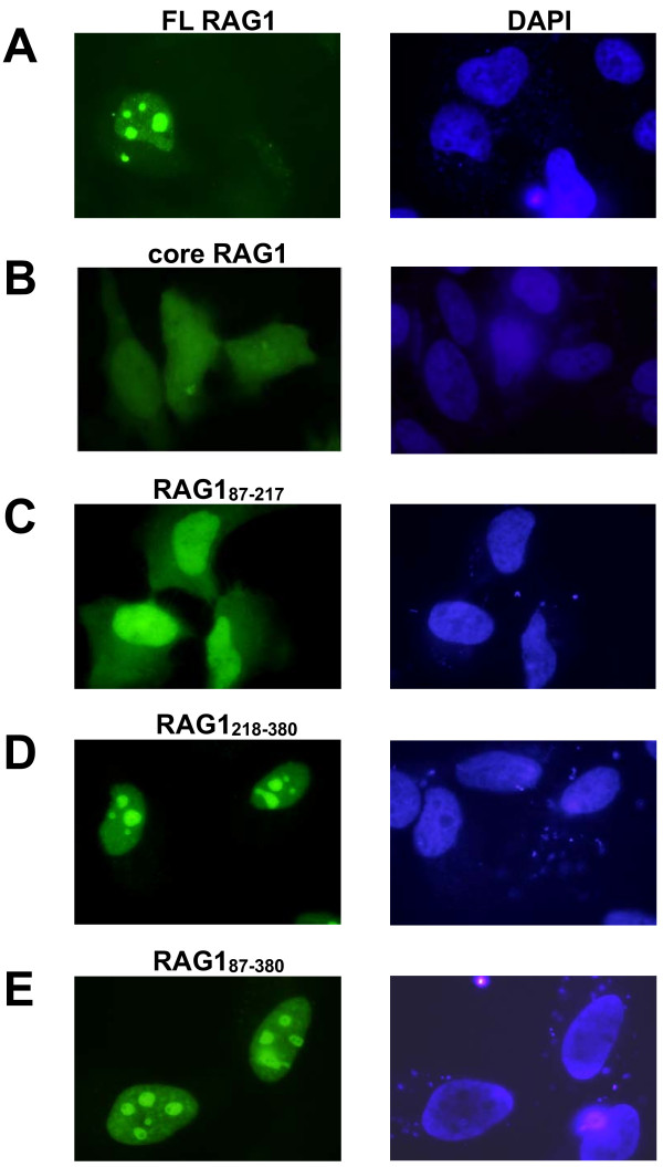
Figure 6.
Nuclear localization of GFP-tagged non-core RAG1 domains. HeLa cells were transfected with GFP fused to the N-terminus of the indicated RAG1 polypeptides. Following 24-48 hrs cell growth, cells were fixed and stained with DAPI. Left panels: fluorescence microscopy images of HeLa cells expressing (A) GFP-full length RAG1, (B) GFP-core RAG1, (C) GFP-CND (residues 87-217), (D) GFP-bZDD (residues 218-380), and (E) GFP-(CND+bZDD) (residues 87-380). Right panels: The corresponding nuclei of cells in the left panels stained with DAPI.

