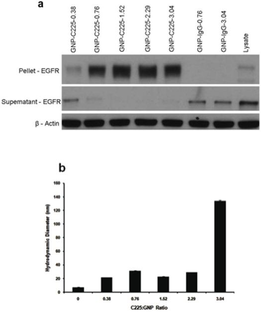
Figure 4. Characterization and in vitro uptake of GNP-C225.
a. Binding of GNP-C225 and GNP-IgG to EGFR verified by Western Blot analysis. Cell lysates from AsPC-1 cells were incubated for 2 hrs at room temperature with GNP-C225 and GNP-IgG conjugates with various ratios of antibody on the surface of the particle. After incubation the samples were spun at high speed and the supernatant was collected and the pellet was washed once and respun; the nanoconjugate fraction was 20 times the concentration of the supernatant. The pellet and the supernatant fractions were loaded on a 7.5% SDS-PAGE gel and analyzed for EGFR. b. Hydrodynamic diameter of GNP-C225 measured by Dynamic Light Scattering Spectroscopy.

