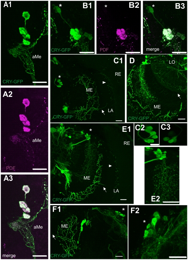
Figure 1. Localization of CRY-positive cells in the brain of Drosophila melanogaster.
Flies were examined at four ZTs: ZT1, ZT4, ZT13 and ZT16 (ZT0 – the beginning of the day and ZT12 – the beginning of the night) but images shown in the Fig. 1 were obtained from the brain of individuals collected for experiments at different ZTs. ZT for each image is given in brackets. A1–3: Four large ventral lateral neurons (l-LNvs), four small LNvs (s-LNvs) and an arborization in the accessory medulla (aMe) immunoreactive to PDF (magenta) and labeled with cry-GAL4-driven GFP (green) (ZT1). B1–3: Double-labeling of the LNvs with PDF antiserum (magenta) and cry-GAL4-driven GFP (green) (ZT16). The 5th s-LNv expresses GFP but not PDF (asterisk). C1: cry-GAL4-driven GFP expression in the optic lobe: LNvs and LNds (asterisk). The LNvs send processes to the medulla (ME) and a single projection to the lamina (LA) which divides and terminates in the lamina cortex (arrows) (ZT4). C2–3: CRY-positive dorsal lateral neurons (LNds). Out of 6 LNds (C2) (ZT4), 3–4 cells have a higher (by 45–80%) intensity of GFP than other LNds in most preparations (C3) (ZT13). D: CRY-positive network of processes in the lobula (LO) (arrow). Processes from DN3s and LNvs in the medulla, also shown. E1–2: Dorsal neurons DN3s and LNvs. The LNvs project to the medulla (ME) and to the lamina (arrows) (ZT4). DN3s form a cluster of cells (asterisk) and send processes to the medulla (E2). F1–2: Network of processes of LNvs with varicosities in the medulla and in the lamina (arrow); CRY-positive DN1s (asterisk, F2) (ZT1). Scale bars: 20 µm; in C2: 10 µm.

