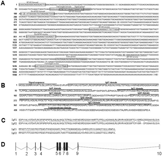
Figure 1. Analysis of cDNA and the protein sequence of porcine 4Ig-B7-H3.
A, The cloned cDNA. The boxed, underlined and gray colored sequences show the positions of all used primers. The numbers in the brackets are the exon order of porcine B7-H3 in the genome. The strong ATG and TGA are initiation and stop codons. All exons contain the complete sequence in the genome but exon 1 in the cDNA. The numbers on the left are the base count of cDNA. B, The predicted protein sequence. The numbers on the left are the count of amino acids . All predicted domains are marked above the sequence. C, Alignment of the duplicated amino acid sequences of the two IgV and IgC-like domains, respectively. Asterisks indicate the identical amino acids. D, Schematic structure of CD276 nucleotide sequence in the porcine genome. The filled panes and lines represent Exons and Introns, respectively. The numbers under the schematics is the order of the Exons in the genome.

