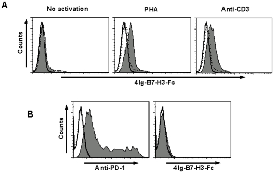
Figure 3. Analysis of a putative receptor of porcine 4Ig-B7-H3.
A, Binding of porcine 4Ig-B7-H3-Fc to T cells. T cells treated in different conditions were stained with a combination of anti-CD3 mAb with Fc (solid line histogram) or isotypic Ab (dotted line histogram) or 4Ig-B7-H3-Fc (filled histogram). Left plot: T cells in PBMC were not activated. Middle plot: T cells in primary PBMCs were stimulated with 5 µg/ml of PHA for 24 h. Right plot: T cells in primary PBMCs were stimulated with 1 µg/ml of anti-porcine CD3 mAb coated on the 96-well plate for 48 h. B, PD-1 gene was transfected into 293 T cells. PD-1 expressed on the 293 T cells was stained with anti-PD-1 mAb (left, filled histogram) or 4Ig-B7-H3-Fc protein (right, filled histogram) or Fc (solid line histogram) or isotypic Ab (dotted line histogram) 24 hours later.

