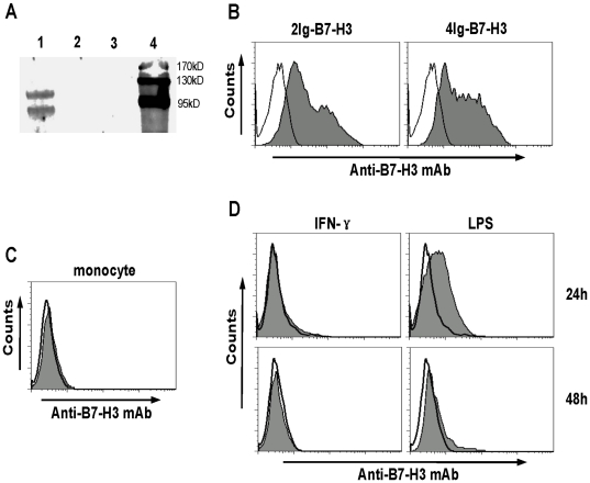
Figure 5. Analysis of porcine B7-H3 expression on monocytes.
A, Immunoprecipitation assay. The membrane protein extracted from the porcine 4Ig-B7-H3-transfected 293 T cells (Lane 1) or vacant plasmid-transfected 293 T cells (Lane 2) was pulled down with anti-porcine B7-H3 mAb-bound protein-G agarose. The pulled-down proteins were submitted to western-blot assay. Lane 3: anti-porcine B7-H3 mAb-bound protein-G agarose control. Lane 4: protein marker. B, The analysis of the porcine 2Ig-(left plot) and 4Ig-B7-H3 (right plot) expressed on 293 T cells stained by the anti-porcine B7-H3 mAb (filled histogram) or isotypic Ab (open histogram). C, non-activated monocytes were stained with a combination of anti-CD14 with either anti-B7-H3 mAb (filled histogram) or isotypic Ab (open histogram). D, The monocytes were co-cultured with IFN-γ in a final concentration of 30 ng/ml (left plots) or LPS at a final concentration of 1 µg/ml (right plots) for 24 h (upper plots) or 48 h plots (lower plots). On the indicated time points, the monocytes were harvested to be stained with a combination of anti-CD14 with either anti-B7-H3 mAb (filled histogram) or isotypic Ab (open histogram).

