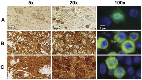
Figure 1. MAGE-C1/CT7 protein expression pattern in melanoma.
MAGE-C1/CT7 expression was analyzed by immunohistochemistry on melanoma lesions at two different magnifications (5x, 20x) (left and middle panels) and by immunofluorescence on cell lines (100x) (right panels). Expression of MAGE-C1/CT7 is shown only in the nucleus (A), only in the cytoplasm (B) and both in nucleus and cytoplasm (C).

