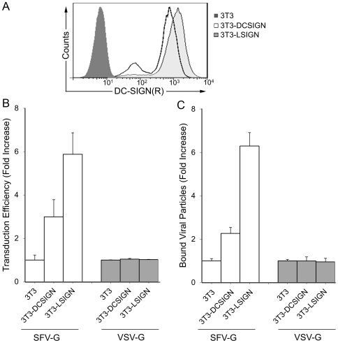
Figure 3. Effects of DC-SIGN or L-SIGN expression.
(A) Expression of L-SIGN and DC-SIGN in 3T3 (solid fill), 3T3.DCSIGN (open fill) and 3T3.LSIGN (gray fill), respectively, was detected using cross reactive DC-SIGN/L-SIGN antibody and quantified by flow cytometry. (B) Effects of DC-SIGN or L-SIGN expression on the infectivity of pseudotyped lentiviruses. SFV-G- and VSV-G-pseudotyped lentiviruses were normalized by p24 and spin-inoculated with LSIGN- or DCSIGN-expressing 3T3 cells; the parental 3T3 cells were included as controls. Three days later, the transduction efficiency was measured by analyzing GFP expression. Fold increase in percentage of GFP-positive cells is shown based on 3T3 cells where FUGW/SFVG transduced 6.6±0.7% and FUGW/VSVG transduced 72.2±1.0%. (C) Specificity of binding to DC-SIGN. [35S]-methionine-labeled FUGW/SFVG or FUGW/VSVG were incubated with 3T3 or DC-SIGN/L-SIGN-expressing cells at 4°C. Cells were washed and 35S radioactivity of the resuspended cells was quantitated with a liquid scintillation counter. Fold increase in [35S] bound viral particles is shown based on 3T3 cells with 3.59±0.27% and 25.23±1.25% of the total CPM of virus bound for FUGW/SFVG and FUGW/VSVG, respectively, where values are given as the mean of triplicates ± S.E.

