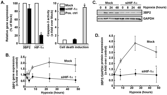
Figure 2. Gene silencing of HIF-1α antagonizes the effects of hypoxia on 3BP2 gene and protein expression.
MSC were transiently transfected with scrambled sequences (Mock, white bars, open circles) or HIF-1α siRNA (black bars, closed circles) as described in the Methods section. Cells were then cultured under normoxic or hypoxic culture conditions as described in the legend of Fig. 1. (A) Apoptotic cell death was assessed using the fluorometric caspase-3 activity assay as described in the Methods section. Concanavalin-A-treated MSC (grey bars) was used as a positive inducer of caspase-3 [16]. Total RNA was extracted, and qRT PCR was used to assess 3BP2 and HIF-1α transcript levels. (B) 3BP2 gene expression was assessed by qRT-PCR in Mock-transfected and in siHIF-1α-transfected cells that were subsequently cultured under hypoxic conditions. (C) Mesenchymal stromal cells were transiently transfected with scrambled sequences or HIF-1α siRNA as described in the Methods section. Cells were then cultured under normoxic or hypoxic culture conditions, cell lysates were isolated, western blotting and immunodetection was performed with anti-3BP2 and anti-GAPDH antibodies. A representative blot is shown out of two. (D) Scanning densitometry was used to assess protein expression described in panel C, and the ratio of 3BP2/GAPDH expression was represented. Values in (A) and (B) are means of two independent experiments, each performed in triplicates (*p<0.05 versus mock control in (A) or mock at time = 0 hr hypoxia in (B)); Bars, ±SD.

