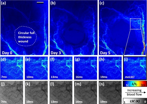
Figure 5.
Use of meLSCI for imaging the microvascular environment during wound healing in a mouse ear injury model: (a–c) The top panel shows sequentially obtained meLSCI images of the mouse ear vasculature of the day of wounding, and three and five days after wounding. Note that the meLSCI scheme provides the contrast necessary to image the changes associated with neovascularization. (d–i) The middle panel shows high magnification views of the boxed region in (c), obtained using LSCI at single-exposure times and using meLSCI. 1/τc values have been depicted in pseudocolor. meLSCI can be clearly seen to have a higher contrast-to-noise ratio in proximal angiogenic regions by comparing (i) with (d–h). (j–n) The bottom panel shows equivalent high magnification views of the boxed region in (c), as they appear when the speckle contrast K is displayed in grayscale. Note that images (j–n) have been linearly and uniformly scaled for better print reproduction. Scale bar shown in (a) corresponds to 0.5 mm.

