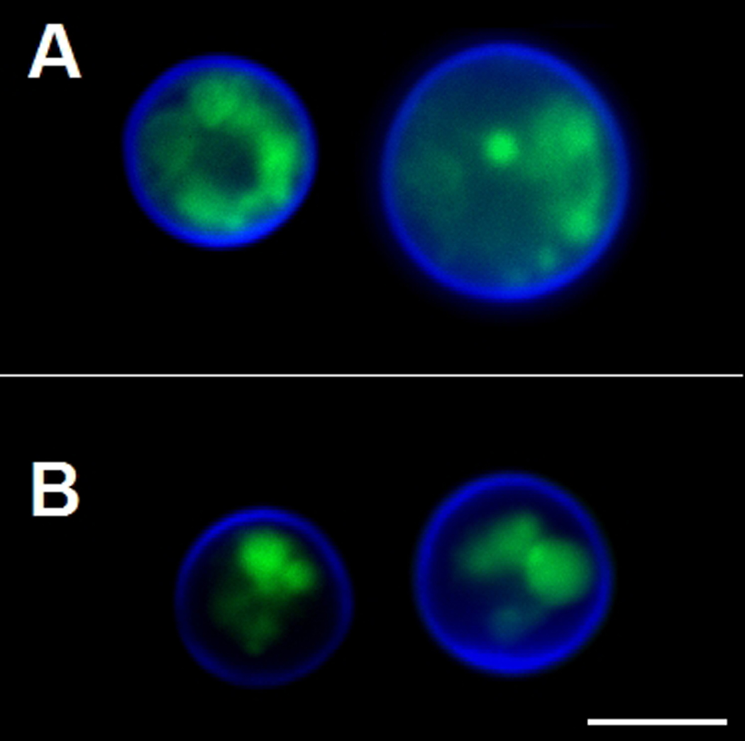
Figure 3.

Modified morphology of the Golgi apparatus in C. neoformans GRASP ortholog mutant cells. WT cells (A) and the GRASP ortholog mutant (B) were sequentially incubated with C6-NBD-ceramide for Golgi visualization (green) and Uvitex 2B for staining of the cell wall (blue). Scale bar, 3 µm.
