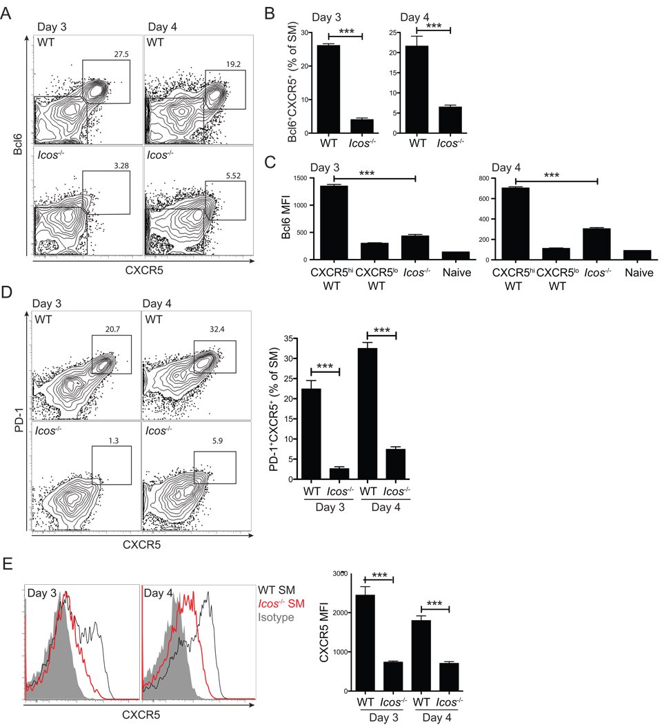
Figure 3. Early Tfh cell commitment requires ICOS signals for Bcl6 expression.
WT or Icos−/− SM CD4+ T cells were transferred into B6 mice subsequently infected with LCMV. (A, B) Bcl6+CXCR5+ Tfh cell development was analyzed 3 and 4 days p.i. by FACS (A) and quantified (B). (C) Bcl6 protein expression was quantified in WT CXCR5+ SM and WT CXCR5 SM vs. Icos−/− SM CD4+ T cells. (D) PD-1 expression on WT and Icos−/− SM CD4+ T cells. (E)Left, CXCR5 expression on WT (black) and Icos−/− (red) SM CD4+ T cells, compared to isotype control (gray filled). Right, CXCR5 MFIs. Data are representative of three independent experiments; n = 4 per group. ***P<0.001.

