Fig. 2.
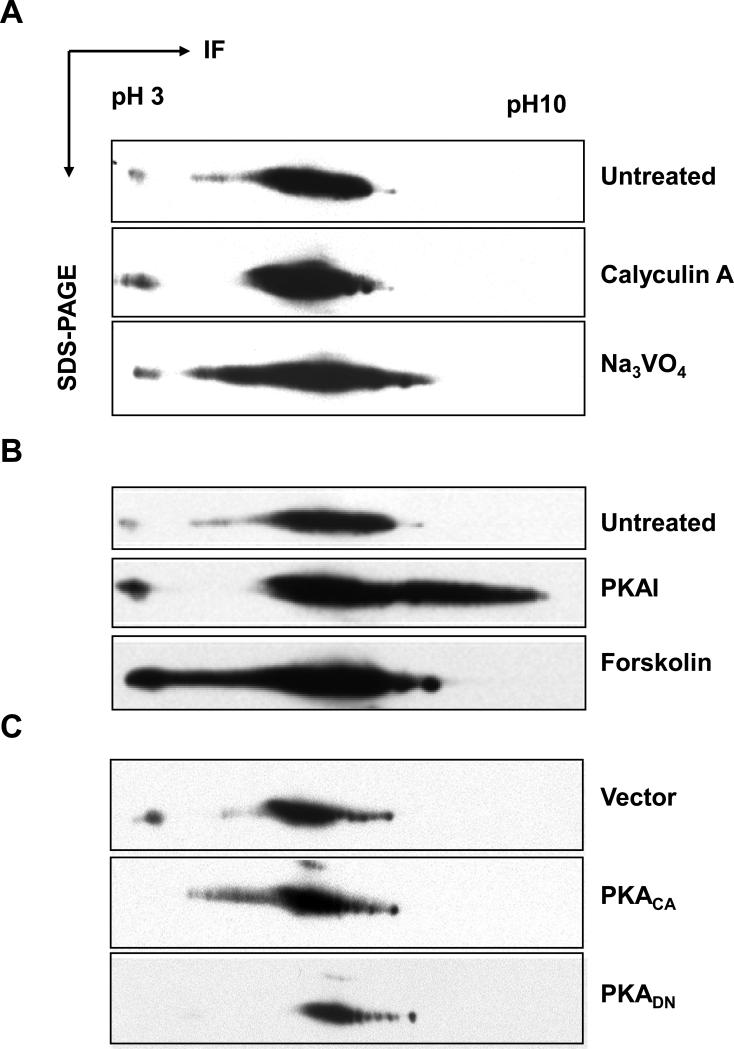
Phosphorylation of SMN in vivo. (A) HEK293 cells were left untreated or treated with calyculin A (threonine and serine phosphatase inhibitor) and sodium vanadate (tyrosine phosphatase inhibitor). Lysates from untreated or treated cells were separated by two-dimensional SDS-PAGE, and the SMN protein (~37 kD) was detected by western blotting using anti-SMN antibodies. (B) HEK293 cells were left untreated or treated with PKA agonist forskolin or PKA inhibitor KT5720. Lysates from untreated or treated cells were analyzed as in A. (C) HEK293 cells were transfected with vector alone, PKA catalytic (PKACA), or dominant negative (PKADN) constructs. Lysates from vector or PKA constructs-transfected cells were analyzed as in A. IF = isoelectrophoresis focusing.

