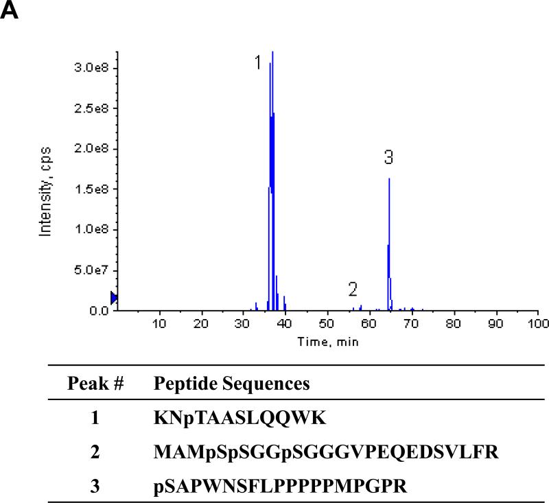
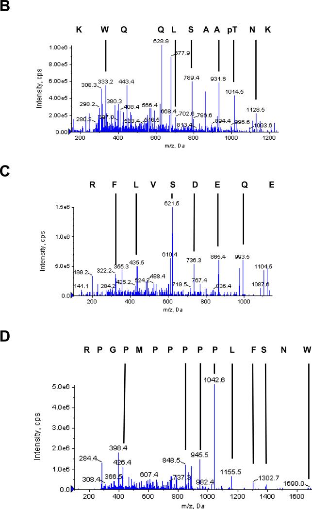
Fig. 4.
MS/MS analysis of phosphopeptides from SMN treated with PKA. (A) Three phosphopeptides were identified by MS of eluted peptides in negative ion mode and the extracted ion chromatogram of the identified peptides is shown. MS analysis of these peptides in positive ion mode identified three phosphopeptides from SMN with a confidence interval ≥ 95%. The corresponding phosphopeptide sequences are shown below. (B-D) Sequences of the three identified phosphopeptides were determined by MS/MS in positive ion mode. Singly charged Y-series peptide ions are identified by masses and vertical lines. The corresponding peptide sequence in C – N direction is shown above the ion spectrum. The ion masses of the parent peptides correspond to mono (peaks 1 and 3) and tri (peak 2) phosphopeptides. Note that the masses of the fragments that contain serine but not threonine correspond to non-phosphorylated peptides, while fragments that contain both the serine and the threonine correspond to modified peptides with loss of one H3PO4. Mascot-based ion assignment is provided in Figure S2.


