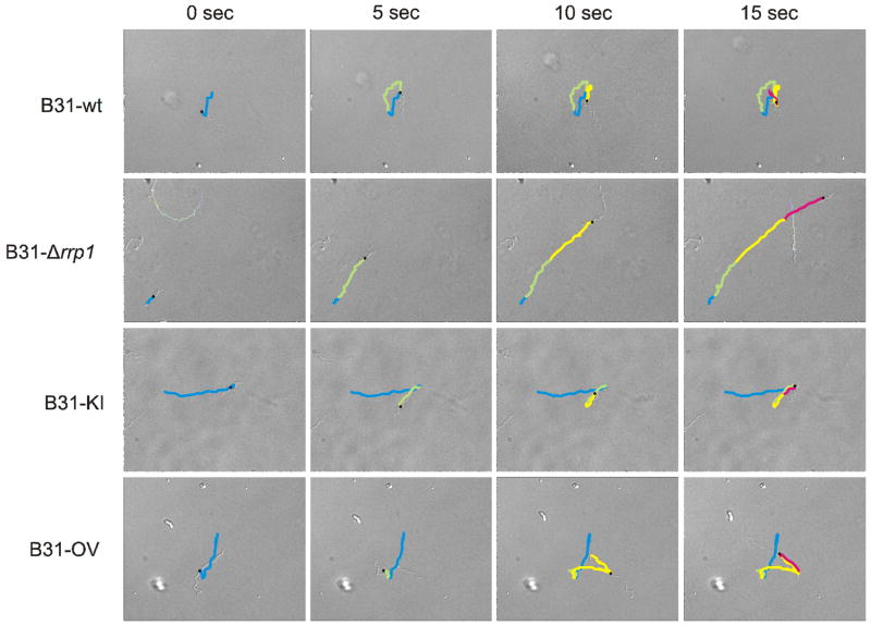
Figure 4. Translational motion patterns requires Rrp1.
Movement patterns of the B31-wt, B31-Δrrp1, B31-KI, and B31-OV strains were visualized using DIC microscopy. Images were captured at 5 sec intervals are shown. Motion tracks were manually recorded and overlaid on the images using motion tracking software. The tracks are colored according to motion achieved during each timelapse (blue - 0 sec, green - 5 sec, yellow - 10 sec, pink - 15 sec). Note that the patterns shown are representative of the entire population of cells.

