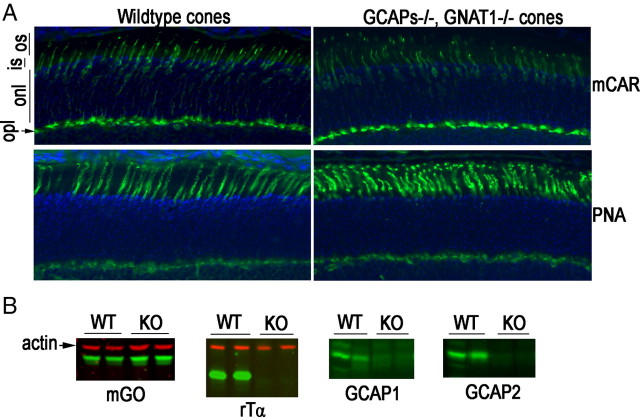
Figure 1.
Cone density and expression of transduction proteins in wild-type and GCAPs−/−,Gnat1−/− retina. Cones were visualized by immunofluorescence (green) (A) of mouse cone arrestin (mCAR, top) and peanut agglutinin (PNA, bottom). Nuclei were stained with DAPI (blue). os, Outer segment; is, inner segment; onl, outer nuclear layer; opl, outer plexiform layer. B, Representative Western blots of whole retinal homogenate from wild-type (WT) and GCAPs−/−, Gnat1−/− (KO) mice probed with antibodies against the indicated transduction protein (mGO, rTα, GCAP1, and GCAP2). Actin served as a loading control. Each lane contains retinal homogenate from a different mouse. No changes in expression level of mouse green opsin were observed.

