
Figure 1.
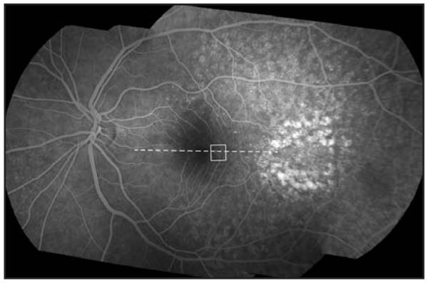
Fundus fluorescein angiography montage of the patient’s left eye. Individual images were montaged using i2k Align Retina software (DualAlign, LLC, Clifton Park, NY). The white box indicates the area of the cone mosaic displayed in Figure 2 and the dashed line indicates the location of spectral domain optical coherence tomography scan shown in Figure 2.