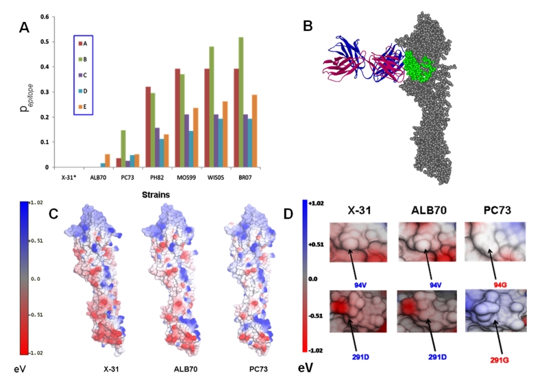
Figure 1.
(A) Antigenic distances of all the epitopes (A, B, C, D and E) on HA proteins from selected vaccine strains of Influenza A/H3N2 computed with X- 31 as a standard. (B) The docking of BH151 Mab into the E epitope of ALB70 HA protein. The VH and VL of the Bh151 are indicated in magenta and indigo colors respectively in Ribbon mode display, whereas, the HA protein of ALB70 is displayed in spacefill mode in Grey color with the epitope E highlighted in Green. (C) The comparison of Surface electrostatics (outputs from NOC program) reveals similarities between the X-31 and ALB70 whereas considerable differences existed between X-31 and PC73. (D) Figure highlighting the altered surface electrostatics in HA protein from PC73 due to mutations V94G and D291G w.r.t ALB70 (and X-31). N.B. The amino acid numbering followed in the text is in accordance with the sequence nomenclature of Influenza A/X-31/1968 HA protein (SwissProt id: P03438).

