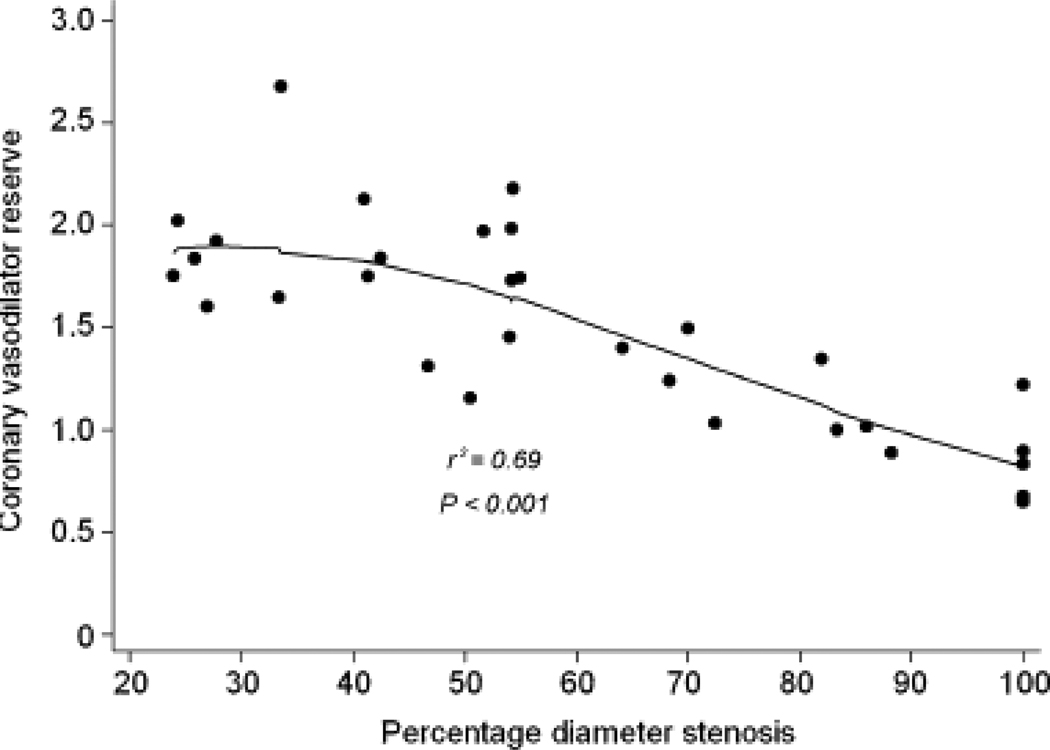
FIGURE 3.
(A) Median values of coronary vasodilator reserve with IQ range (25–75percentiles) and with upper and lower adjacent values; *p value from Kruskal Wallis analysis for four groups (<50%, 50%–69%, 70%–89% and >90% diameter stenosis). (B) Scatter plot of relation of coronary vasodilator reserve and quantitative coronary angiography measurements of percent diameter stenosis.

