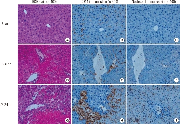
Fig. 4.
Hepatic histopathology following I/R. Mice were subjected to 60 min of ischemia followed by reperfusion at the time point shown. The ischemic liver sections were prepared and stained with H&E and then immunohistochemical staining of CD44 and neutrophils using specific anti-CD44 monoclonal antibody and anti-neutrophil antibody. (A), (B), and (C) represent the sham mice. There are normal hepatic histology (A). Nornal liver lobule shows mildly positive CD44 immunostaining (B) and negative neutrophil immunostaining (C). (D), (E), and (F) represent the mice subjected to 60 min of ischemia followed by reperfusion for 6 hr. The hepatic lobules show one focus of coagulation necrosis and adjacent ballooning degeneration (D). The liver shows moderately positive CD44 immunostaining (E) and focal a few positive neutrophil cells (F). (G), (H) and (I) represent the mice subjected to 60 min of ischemia followed by reperfusion for 24 hr. There are several large areas of coagulation necrosis and neutrophilic infiltration (G). The liver shows obiously patchy increased CD44 immunostaining (H) and neutrophil influx (I).

