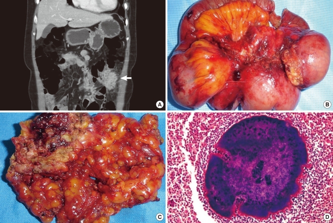
Fig. 1.
Fifty eight years old female presented with lower left quadrant pain for 15 days. (A) Ovoid mass of sigmoid colon mimics a malignant tumor on computerized tomography. (B) 10.0 × 8.0 cm ovoid mass on the sigmoid colon and diverticulitis with severe inflammatory adhesion to descending colon and mesentery. (C) The cut surface demonstrates typical abscess with yellowish-brown color. (D) A magnified view of the characteristic sulfur granule in the middle of purulent exudates (H&E, × 200).

