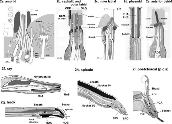
Figure 2.
Ultrastiucture of C. elegans cilia. 2a. Cilia in the amphid sensillum exhibit a variety of morphologies. The rod-like channel cilia are found in ASE, ASG, ASH, ASI, ASJ, ASK, ADF, and ADL neurons. ADF and ADL possess two cilia each, while the other cells possess a single cilium. These cilia are exposed to the environment through the cuticle. The amphid wing neurons (AWA, AWB, AWC) have complex ciliary structures. The AFD neuron possesses multiple villi. The sheath and socket cell encapsulate the amphid channel cilia, whereas the sheath cell encloses the amphid wing (AWA/AWB/AWC) and AFD cilia. Cross section views are shown on the left (a–e). This is modified from (4), Copyright (1986), with permission from Elsevier. . 2b. The cephalic and outer labial sensillum. In the male, the CEM neuron is also located in the cephalic sensillum. 2c. The inner labial sensillium contains IL1 and IL2 neurons. IL2 is embedded within subcuticular structure, while IL1 is exposed to the exterior. 2b and 2c were modified from (1) Copyright (1975, Ward et al) with permission of Wiley-Liss, Inc., a subsidiary of John Wiley & Sons, Inc. 2d. The phasmid sensillum in the tail. The PHA and PHB cilia extend in parallel to each other. Two phasmid socket cells (socket 1 and 2) surround the sensillum. 2e. The anterior deidrid ADE cilia. The image was provided by Sam Ward. 2f. Each ray sensillum contains a pair of cilia: RnA and RnB. RnA cilia are embedded whereas RnB (except R6B) cilia are exposed. The ray structural cell functions as both socket and sheath cell. 2g. The hook sensillum encloses HOA (embedded) and HOB (exposed). 2h. The spicule sensillum has SPV and SPD ciliated neurons, four syncytial socket cells, and two syncytial sheath cells. 2i. The postcloacal sensillum (p.c.s.) possesses one ciliated neuron (PCA), which ciliary tip ends within the cuticle. 2d and 2f–2i. Reproduced from (6) Copyright (1980), with permission from Elsevier.

