
Figure 6.
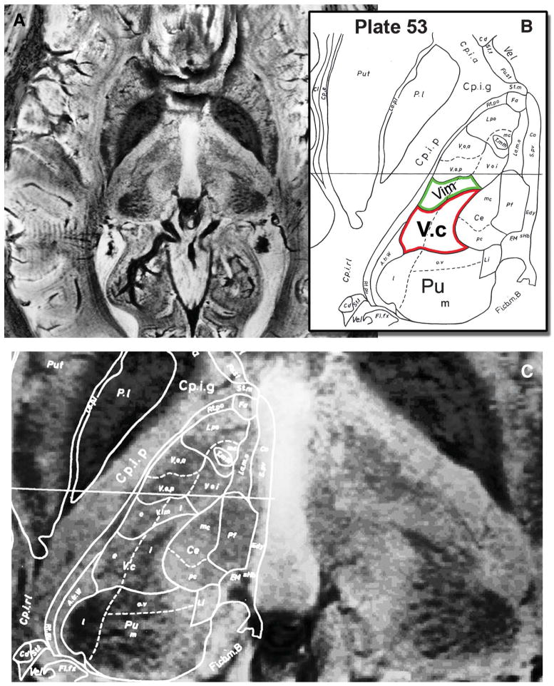
Direct visualization of internal thalamic nuclei in vivo. A, an axial susceptibility-weighted imaging slice through the thalamus at the level of the anterior and posterior commissure plane is shown. B, the corresponding histologically defined outline (plate 53 from the Schaltenbrand and Wahren atlas18) that is superimposed on the magnetic resonance image in C. Note the clear visualization of the anterior and medial aspects of the pulvinar (Pu), the arrowhead shape of ventral caudalis (Vc; red outline; compare with unmarked right hemisphere), and the image contrast modulation within the thalamus corresponding to Vim (green outline).