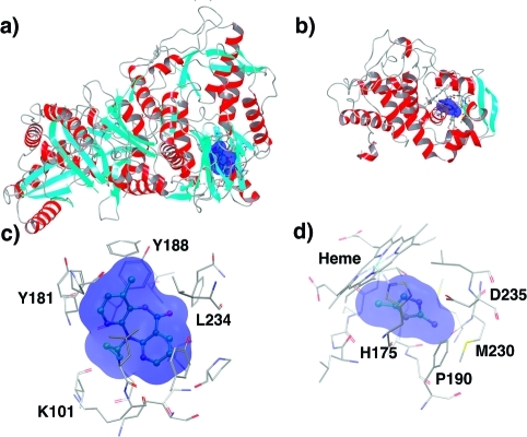
Figure 1.
Protein receptors considered in this study: (a) RT and (b) W191G overall representations on the same scale. Secondary structure elements and the location of the binding sites are highlighted (red: helices; cyan: sheets; and gray: loops and turns). Insight views for: (c) the RT NNRTI binding pocket (NNIBP) with nevirapine bound and (d) the W191G cation-binding pocket with 2a5mt bound. Ligands (balls and sticks) and pocket volumes (blue surfaces) are also shown.

