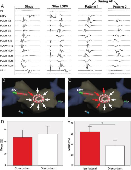
Figure 5. Organized patterns at the posterior left atrial wall.
A. Left, activation patterns during sinus rhythm and during stimulation from the left superior pulmonary vein (LSPV). Right, two activation patterns were observed during atrial fibrillation (AF): pattern 1 resembled LSPV stimulation; pattern 2 was similar to sinus rhythm activation. B and D. During AF, concordant patterns of activation resembling LSPV stimulation from which AF was induced (red arrow originating at the pacing catheter) accounted for 46±27% of recording time when compared discordant patterns (white arrows). C and E. Organized patterns during AF resembling those obtained during pacing from any of the ipsilateral PVs (red arrows originating from the pacing PV side, superior and inferior) accounted for 63±24% of the organized time (p=0.025). CSd, distal coronary sinus.

