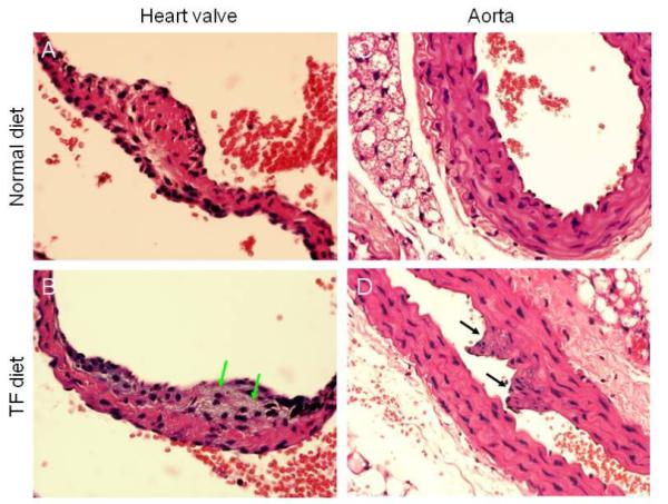
Fig. 2. Histological analysis of heart and aorta in mice fed TF and control diets.

Mice were fed TF and control diets for 24 weeks. Heart valves (A, B) and aorta (C, D) were analyzed following fixation and H & E staining. A representative of a total of 5 mice analyzed is shown. Foam cells and fibro-intimal proliferation were found in the heart valves and aorta as indicated by green and black arrows, respectively.
