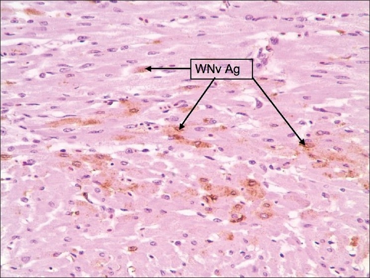
Figure 6.

Heart: Staining was faint, diffuse, and scattered. Stained cells were macrophages, myofibers, and occasionally endothelial cells. Pericardium showed sections of intense staining of macrophages

Heart: Staining was faint, diffuse, and scattered. Stained cells were macrophages, myofibers, and occasionally endothelial cells. Pericardium showed sections of intense staining of macrophages