Figure 5.

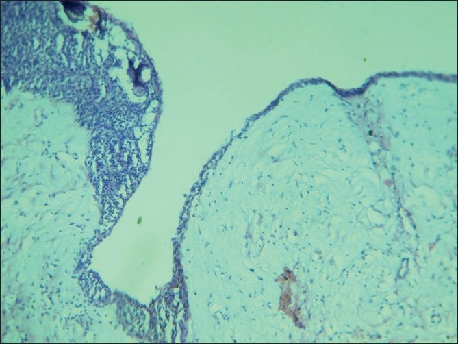
10× view showing a thin nonkeratinized lining epithelium continuous and showing proliferation in one area, spindle cells and calcifications are seen

10× view showing a thin nonkeratinized lining epithelium continuous and showing proliferation in one area, spindle cells and calcifications are seen