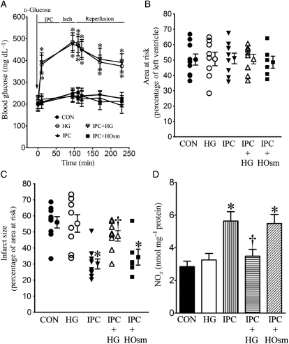
Figure 2.
HG inhibited decreases in myocardial infarct size produced by IPC in WT mice subjected to I/R injury. (A) Blood glucose concentrations during myocardial ischaemia (Isch) and reperfusion; (B) AAR expressed as the percentage of the left ventricle; (C) infarct size expressed as the percentage of AAR. All mice were subjected to 30 min of coronary occlusion followed by 2 h of reperfusion (CON). IPC was induced by four cycles of 5 min of ischaemia/5 min of reperfusion. Mice were injected with d-glucose to produce HG or mannitol to produce hyperosmolarity (HOsm); (D) NO concentrations. The hearts were collected 5 min after reperfusion, and myocardial NO and its metabolites (nitrate and nitrite) were measured. *P < 0.05 vs. CON, †P < 0.05 vs. IPC (n = 6–10 mice/group).

