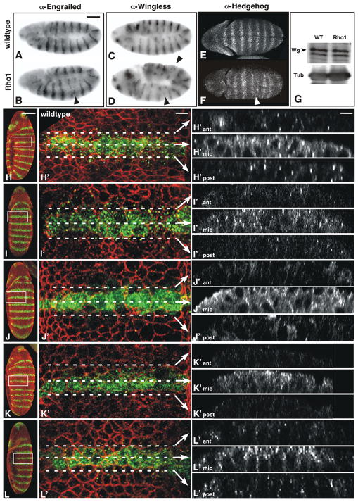
Figure 1. En and Wg expression is not maintained properly and Wingless protein distribution is aberrant in maternal Rho1 mutants.
Stage 9 wildtype (A, C, E) or maternal Rho1 mutant (B, D, F) embryos stained with anti-Engrailed (A–B) anti-Wingless (C–D) or anti-Hedgehog (E–F) antibodies. While En expression is initiated normally, as development proceeds some stripes become reduced, others disappear entirely (arrowhead in B). Likewise, early Wg and Hh expression in maternal Rho1 mutants looks normal, but in later stages Wg and Hh stripes also become reduced or eliminated (arrowheads in D, F). (G) Western analysis of Wg protein levels in wildtype and maternal Rho1 mutant embryo lysates. Tubulin levels shown as loading control. (H-L′) Z-series projections of wildtype (H, H′), wimp/+ (I, I′), shi (J, J′), maternal Rho1 mutant (K, K′), and maternal Rho1 rescue (L, L′) stage 9 embryos stained with antibodies against Wingless (Wg, green) and Discontinuous Actin Hexagon (Dah, red) to outline cell boundaries. The boxed regions of embryos in H, I, J, K, L are shown at higher magnification in H′, I′, J′, K′, L′. Z-series cross-sections noted by dashed lines in H′, I′, J′, K′, L′ are projected in ant, mid, post. Note the punctate accumulations of Wg protein spreading out from the Wg-expressing cells in wildtype and wimp/+ (H-I′), which are reduced in shi mutants (J-J′), which are defective in endocytosis, and maternal Rho1 mutants (K-K′). This phenotype is rescued by the presence of a Rho1 transgene (L-L′). The larger cells in J′ relative to H′ result from cytokinesis defects associated with shibire mutants (Bejsovec and Wieschaus, 1995). Scale bars: (A, H) 100μm; (H′) 10μm.

