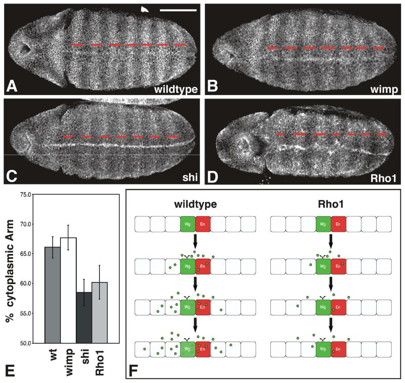
Figure 4. Maternal Rho1 mutants exhibit aberrant cytoplasmic Armadillo accumulation.
(A-D) Representative stage 9 wildtype (A), wimp (B), shibire (C), or maternal Rho1 mutant (D) embryos stained with antibodies to Arm. (E) Graph depicts the percentage of the embryo length between stripes 2–7 that consists of cells with elevated levels of cytoplasmic Arm (red dashes in A–D) for each of the genotypes listed above. shibire and maternal Rho1 mutants show decreased overall stripe width relative to wildtype or wimp controls (n=10, p<0.001). (F) Schematic depicting wildtype embryos (left) and maternal Rho1 mutants (right). Wg (green) spreads outward from Wg-expressing cells as development proceeds (top to bottom). Wg is found in the extracellular space and vesicular structures within cells. In Rho1 mutants the number of vesicles is reduced, and the Wg protein gradient does not form properly. Scale bar: 100μm.

