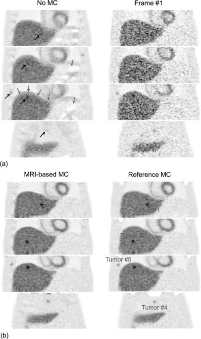
Figure 6.
Simulated PET images in the coronal view reconstructed with (MRI-based MC) and without motion correction (No MC) and using only events from the first frame (Frame #1). Also shown are images corrected using the reference motion fields (Reference MC), which represents the best achievable performance of our method at this noise level. Grey arrows show artifacts on the uncorrected image due to attenuation∕emission mismatches. Black arrows show regions of the uncorrected image with significant motion blurring.

