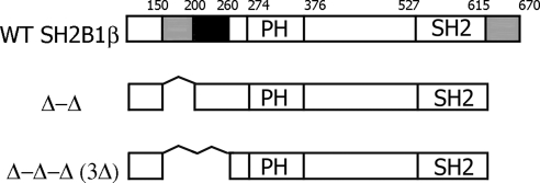
Fig. 1.
Schematic representation of WT and mutant forms of rat SH2B1β used in the study. Actin-binding domains (amino acids 150-200 and 615-670) are shown in gray. Filamin-binding domain (amino acids 200-260) is shown in black. PH is the plekstrin homology domain (amino acids 274-376), and SH2 is the SH2 domain (amino acids 527-620). Proline-rich regions (amino acids 13–24, 89-103, and 469-496) and dimerization domain (amino acids 24-85) are not shown.

