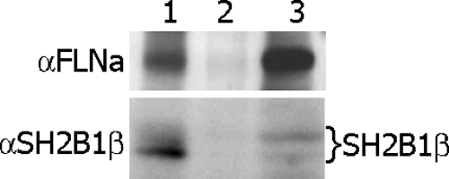
Fig. 6.
Endogenous SH2B1β associates with endogenous FLNa. Whole-cell lysates of 293T cells (lane 3) were immunoprecipitated with αSH2B1β antibody (lane 1) or with control IgG (lane 2) and immunoblotted with αFLNa antibody (upper). Endogenous FLNa was detected in the whole-cell lysate (lane 3) and in the αSH2B1β immunoprecipitate (lane 1) but not the control lane (lane 2). The same blot was reprobed with αSH2B1β (bottom). Several bands representing different levels of SH2B1β phosphorylation (indicated on the right) were detected in the cell lysate (lane 3) and αSH2B1β immunoprecipitate (lane 1) but not in the control lane (lane 2). Each experiment was performed at least three times with similar results.

