Fig. 4.
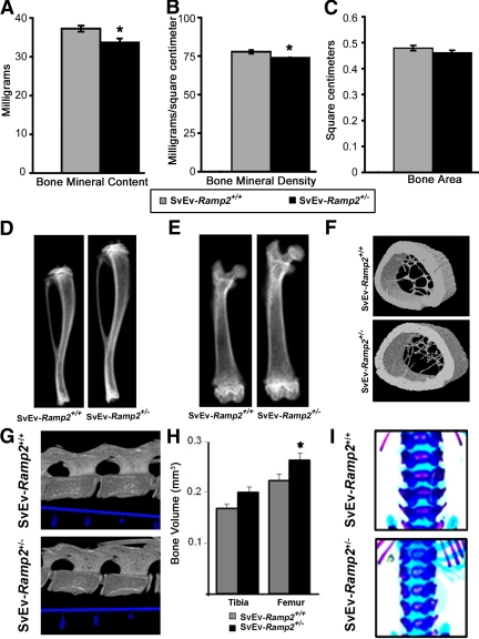
Ramp2+/− mice have skeletal abnormalities. A–C, DEXA analyses of femurs showing decreased bone mineral content in milligrams (A), decreased bone mineral density in milligrams per square centimeters (B) (*, P < 0.01) and C) unchanged bone area in square centimeters (C); D, radiographs of tibiae; E, radiographs of femurs; F, micro-CT of midregion of femurs; G, 3D model of lumbar vertebrae; H, tibial and femoral bone volume (*, P < 0.05); I, skeletal staining by Alcian blue; Alizarin red (unmineralized bone and cartilage in blue, mineralized bone in purple). Animals used for the experiments in A–G are 18-wk-old virgin female SvEv-Ramp2+/+ and SvEv-Ramp2+/− mice; n = 8–10 per genotype for DEXA, and n =3 for micro-CT analysis and radiographs. Animals used in H and I are 2-d-old pups of SvEv-Ramp2+/+ (n = 5) and SvEv-Ramp2+/− genotypes (n = 3).

