Figure 3.
Retrospective tracing of metaplasia through embryogenesis
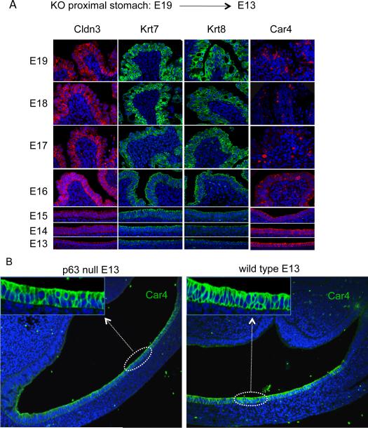
A. Fluorescence micrographs of metaplasia in proximal stomach of p63 null embryos from E19 to E13 stained with antibodies to Cldn3, Krt7, Krt8, and Car4 and counterstained with Hoechst dye for DNA (blue). B. Sections through the proximal stomach of E13 p63 null (left) and wild type (right) embryos stained with antibodies to Car4 (green) showing a simple columnar epithelium lining the lumen. Insets at higher magnification. Sections counterstained with Hoechst dye for DNA. See also Figure S4.

