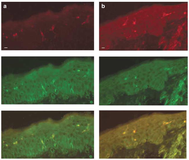
Figure 3. Vα24+/Vβ11+ NKT cells are detected in ACD skin biopsy specimens by immunohistochemistry.
Frozen sections from skin-biopsy specimens of ACD were double-stained with anti-Vα24 and Vβ11. (a and b) Double staining of two different biopsy specimens (CD2 and 4) revealed that all Vβ11 (red, top panel)-bearing T cells also expressed Vα24 (green, middle panel); overlay (orange-yellow, bottom panel) (bar=10 μm).

