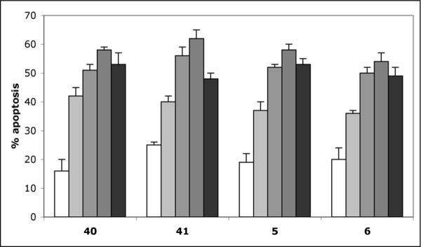
Figure 6.
Induction of apoptosis in Jurkat cells treated with 40, 41, 5 and 6 (all used at 1 μM) for 12 h (open columns), 24 h (light grey columns), 36 h (medium grey columns), 48 h (dark grey columns), 60 h (black columns), determined using the flow cytometric annexin-V/propidium iodide assay. Error bars represent data from four replicates of a single experiment repeated twice with similar results.

