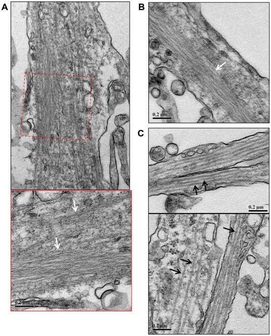
Figure 5. Rod-like accumulations in neurites of primary chick neurons contain densely packed filaments.
Transmission electron micrographs of primary chick neurons treated with 1 µM AM for 15 min, fixed and processed for TEM as described in Methods. (A) Densely packed linear arrays of filaments occur in the neurite (white arrows). The lower panel shows a higher magnification view of the boxed region above. (B) Another example of densely packed filaments within a neurite (white arrow). Here the filament bundle spans a large proportion of the width of the neurite. (C) In contrast, unaffected neurites contain individual separated microtubules approximately 24 nm in diameter (black arrows). Scale bars = 0.2 µm.

