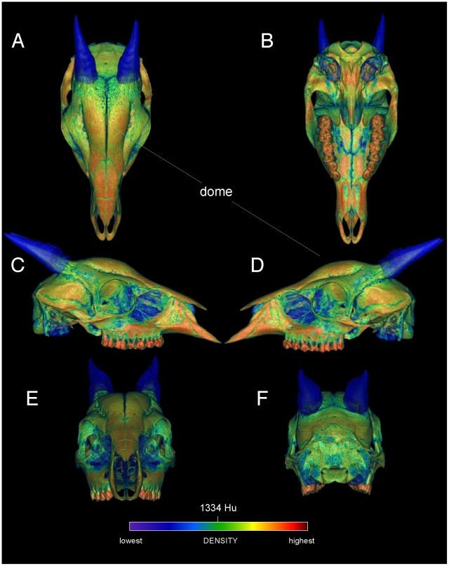
Figure 3. Surface densities of cranial bone in the duiker Cephalophus leucogaster (AMNH 52802).
External cranial densities of the white-bellied duiker, in dorsal, ventral (A, B), right and left lateral (C, D), and anterior and posterior (E, F) views. Duikers collide with a rounded dome formed by thick frontals (the frontals are not fused, as in Stegoceras). The color scale is in Hounsfield units, centered at 1334 (water = 0). The horn sheaths are rendered as slightly transparent, to emphasize high densities of the horn cores; compare with the musk oxen (Figures 4, 5, 6).

