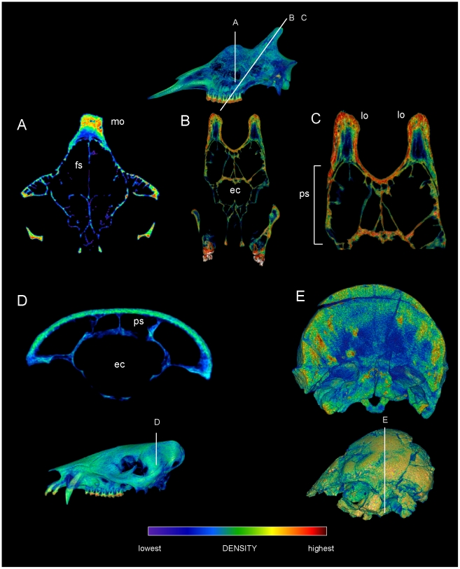
Figure 10. Cranial densities in Giraffa (TMM M 6815, UCMZ 1976.33), the peccary Tayassu (UCMZ 1975.279), and pachycephalosaur Prenocephale (GI SPS, field number PJC2004.8).
CT sections through crania of comparative taxa, with slices mapped onto lateral renders of crania. A. Giraffa camelopardalis male (TMM M6815), transverse section through the region of a median ossicone. B. Oblique transverse section of Giraffa camelopardalis (UCMZ 1976.33) through the posterior ossicones. C. Enlargement of B focusing on the ossicones. The layering of densities in the giraffe ossicones resembles that in the dome of Stegoceras validum (Figure 6). D. Transverse section through the cranium of Tayassu tajacu, showing a non-cancellous skull roof over cranial sinuses. D. Section through the cranium of the pachycephalosaur Prenocephale prenes (GI SPS). Despite mineral inclusions (localized red and yellow areas) and CT artifacts, the scan shows dense superficial and cancellous deep regions within the dome. Abbreviations: ec = endocranial cavity, fs = frontal sinus, lo = lateral ossicone; mo = median ossicone; ps = parietal sinus.

