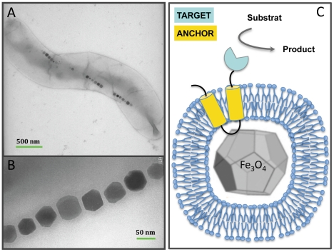
Figure 1. Functionalization of bacterial magnetosomes.
(a, b) TEM image of magnetosomes in Magnetospirillum magneticum AMB-1 cells. The magnetite crystals are aligned within the cytoplasm of the cells on a cytoskeleton made of actin-like proteins. (c) Functionalization of the magnetosome membrane. The targeted enzyme (blue) is anchored on the lipid-coated magnetite crystals by fusion with a membrane protein (yellow).

