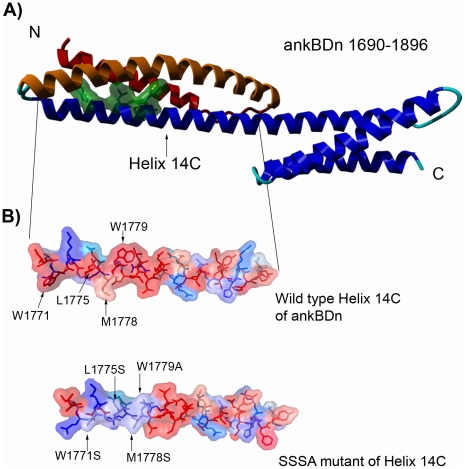
Figure 1. Models of ankyrin-binding domain.
A) Ankyrin-binding domain, with residues W1771, L1775, M1778, W1779 marked in green. Helix 14A is shown in red, helix 14B is colored orange. B) Hydrophobicity analysis of a 38 amino acid fragment of ankyrin-binding domain (ankBDn) and its mutant (SSSA) by 3D-mol software. Colors represent hydrophobicity of amino acid residues: deep red, hydrophobic; and deep blue, hydrophilic. Arrows indicate exchanged residues.

