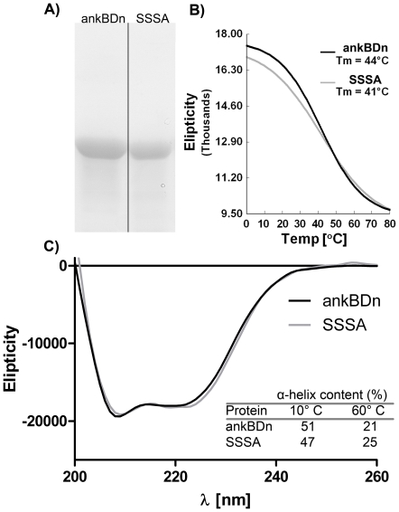
Figure 2. Purity and secondary structure of wild-type and mutant protein.
A) Purified proteins. Electrophoresis was carried out in 10% polyacrylamide gel in the presence of 0.1% SDS. Gels were stained with Coomassie Blue. Arrow indicates ∼42 kDa molecular mass. B) Melting curves of proteins shown in B). The Tm was determined by second derivative calculation. C) CD spectra of recombinant ankyrin-binding domain (ankBDn) and quadruple mutant SSSA at 20°C. Inset: comparison of α-helix content calculated from CD spectra obtained at indicated temperature.

