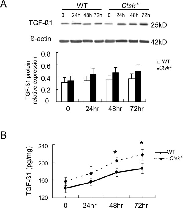
Figure 5.
Effect of CatK expression on TGF-β1 expression and secretion in mouse lung fibroblast (MLFs). (A) shows representative images of immunoblot and band analysis of TGF-β1 expression in WT and CatK-deficient MLFs at indicated time points. (B) shows ELISA analysis of TGF-β1 secretion from MLFs into conditioned media. (* p < 0.05, ** p < 0.01 compared with same time point. All experiments were performed three or four times.)

