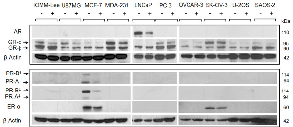
Figure 5.
Expression of progesterone, androgen, glucocorticoid and estrogen receptors in cancer cells exposed to MF. Cells were exposed to either vehicle or the IC50 concentration of MF specific to each cell line for 24 h. Cells were subsequently harvested, lysed, and whole-protein extracts (50 μg for GR and AR; 100 μg for PR and ER-α) were separated by electrophoresis. Immunoblots were then probed with the indicated antibodies (for PR: †clone hPRa7, Thermo Fisher Scientific; ‡ #1483-1, Epitomics); two commercially available antibodies for human PR were used to strengthen the reliability of the results. β-actin was included as a control for protein loading. Because AR, GR-α and GR-β were immunoblotted in one membrane and PR-A, PR-B and ER-α were blotted in a separate membrane, each membrane was blotted separately with anti-β-actin. This experiment was repeated twice with similar outcome. MDA-231 means MDA-MB-231.

