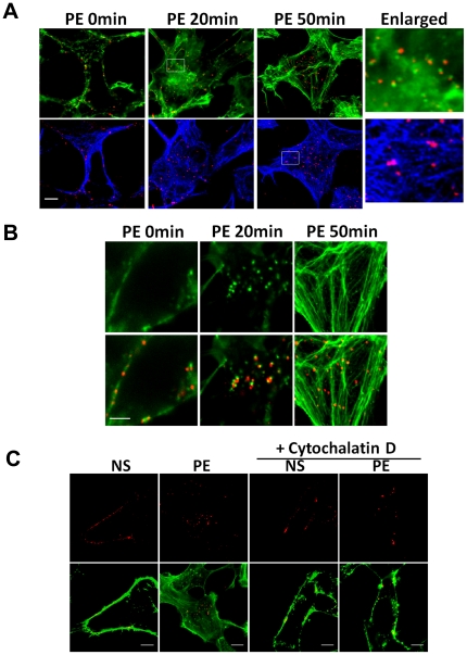
Figure 2. α1A-AR endocytosis is regulated by cytoskeleton.
(A) Colocalization of α1A-AR with F-actin and microtubules after agonist stimulation. Cells were stimulated with 10 µM PE for 20 or 50 min. Untreated cells were used as control. α1A-AR was labeled with anti-FLAG antibodies and Alexa 555 IgG (red). F-actin was labeled with Alexa 488-conjugated phalloidin (green). Microtubules were labeled with antibodies and Alexa 633 IgG (blue). Last column: 5× magnification of selected boxed regions. Bar: 10 µm. (B) High-resolution imaging of colocalization of α1A-AR with reorganized actin after stimulation. Cells were treated with agonist for 20 or 50 min, and then labeled with antibodies against α1A-AR and Alexa 488-conjugated phalloidin against F-actin (red) (bottom row). Bar: 5 µm. (C) Inhibition of α1A-AR endocytosis by Cytochalasin D. HEK-293A–α1A-AR cells were pre-incubated with cytochalasin-D (Cyto-D; 5 µM, 5 min), then stimulated with 10 µM PE for 20 min. F-actin was stained by Alexa 488-conjugated Phalloidin (green), α1A-ARs were detected with anti-FLAG antibody and Alexa-555 IgG (red). NS: no stimulation. Bar: 10 µm.

