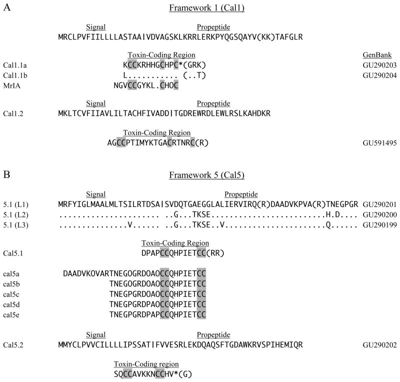
Fig. 1.
Four-Cys sequences of frameworks 1 and 5. In this (and subsequent figures), cDNA clones have the initial species letter capitalized and isolated peptides are designated by all lower case letters (Walker et al., 1999). Vertical bars indicate potential signal peptide cleavage regions, and parentheses show predicted sites of proteolysis in propeptide and mature toxin regions. Predicted C-terminal amidation is indicated by an asterisk (*). A. The framework 1 toxins, Cal1.1a and 1.1b, share a common precursor region (signal and propeptide). Conotoxin Mr1A is shown for comparison. B. For the framework 5 toxin Cal5.1, three leader sequences (L1–L3) were observed. Peptides cal5a–cal5e were identified in venom and correspond to leader sequence L1. O designates hydroxyproline.

