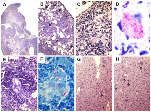
Figure 4. Lung, liver and spleen histopathology of C57BL/6 WT and GKO mice infected with the P104 or H27 M. avium strains.
Representative lung histopathology at 60 days after P104 strain challenge in the WT (B) and GKO (A, C–E) mice and spleen (F) of the GKO mice. Lung presented parenchyma consolidation by epithelioid granulomatous infiltration (A), multifocal lesions with typical granuloma (B - arrows), cellular infiltration containing multinucleated giant cells (C - arrows) with intracellular fuchsinophilic acid-fast bacilli (D); granuloma containing multinucleated giant cells (E) and presence of BAAR inside the intrafollicular granuloma in spleen (F). Representative liver histopathology at 90 days after the challenge of the WT mice with H27 (G) and P104 (H) strains. The tissue samples were stained with H&E (A, B, C, E, G and H) or by Ziehl-Neelsen method (D and F). Total original magnification: A (4x); B, G, H (10x); C, E (20x); F (40x); D (100x).

