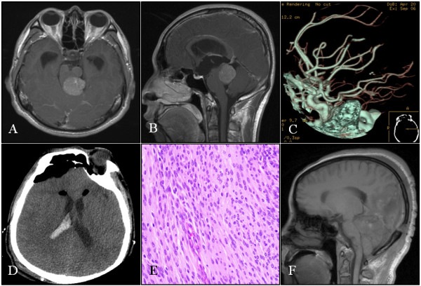
Figure 3.

Case 10: A-B: Axial and sagittal sections of MRI scans that show a contrast-enhanced tumor in the pineal region below the tentorium compressing the brainstem. C: CTA did not demonstrate tumor staining or a feeding artery. D: Postoperative CT scan showing hemorrhage in the lateral ventricle and pneumocephalus. E: Histopathological section of the tumor showing a WHO grade II meningioma (HE, x200). F: Postoperative sagittal MRI section showing no residual tumor.
